Hendra and Nipah Viruses: Catalysing Urgency in Vaccine Development and Diagnostic Advancements
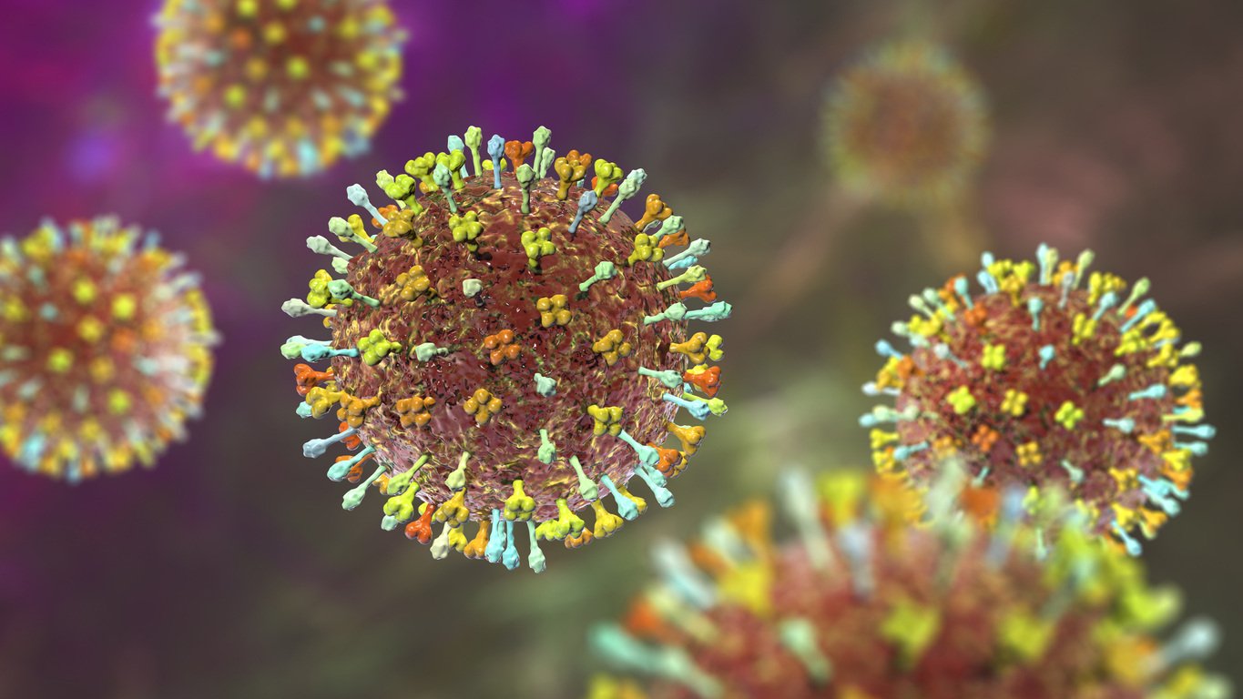
Hendra and Nipah viruses are two closely related pathogens that belong to the genus Henipavirus within the family Paramyxoviridae. These viruses have garnered significant attention due to their zoonotic nature and their potential to cause severe and often fatal diseases. Hendra virus was first identified in Australia in 1994, while Nipah virus emerged in Malaysia in 1999. Both viruses share similar transmission dynamics, primarily involving fruit bats as natural reservoirs and intermediate hosts. They have caused outbreaks with devastating consequences for human health and agricultural economies. With no vaccines available, a wide range of reliable but affordable diagnostics is the only way to control the spread of these deadly viruses.
Sensitive and accessible diagnostics and medication remain the only tools to tame the spread and protect the population from those extremely fatal viruses. At The Native Antigen Company, we're proud to offer a comprehensive selection of Nipah and Hendra virus antigens tailored for vaccine research and efficient immunodiagnostics. By providing these essential tools, we strive to support scientific endeavours to better understand and combat these viruses, ultimately contributing to global health initiatives and the protection of populations worldwide.
The latest additions to our product portfolio include Hendra virus glycoprotein G, Nipah virus Bangladesh, and Malaysia strain glycoprotein G. These glycoproteins are presented tag-free, ensuring cross-reactivity with typical purification handles such as Fc-tags is avoided in your assay design!
Nipah Virus: major threat in South-East Asia
The Nipah virus significantly threatens human and animal health, causing substantial economic losses, particularly in the agricultural sector, where it affects pigs. Human Nipah virus infection is commonly transmitted from fruit bats of the genus Pteropus, commonly known as flying foxes, to humans who drink contaminated raw date palm sap, fruits, and fruit products, as well as through human-to-human contact (Nahar et al., 2015).

Figure 1. Frequently, sap is gathered from date palm trees using a small wooden pipe and collected in clay pots for direct consumption. Bats, notably Pteropus species, are common visitors to these trees and often lick the sap flowing into collection pots from shaved tree parts. These bats can shed NiV through their saliva and urine, potentially contaminating the collected sap (Park, et al 2018).
First identified in a 1999 outbreak among pig farmers in Malaysia, NiV has since caused recurring epidemics in Bangladesh, with sporadic occurrences in eastern India. While Malaysia has not reported new epidemics since 1999, Bangladesh has experienced nearly annual occurrences. Moreover, evidence suggests that other regions may also be at risk due to the presence of NiV in various bat species across several countries, including Cambodia, Ghana, and Indonesia.
Human infections present a broad range of symptoms, from asymptomatic to severe, including acute respiratory infection and fatal encephalitis. Initial signs include fever, headaches, muscle pain, vomiting, and sore throat, progressing to neurological symptoms like dizziness and altered consciousness. Some may develop atypical pneumonia and severe respiratory distress. Severe cases can rapidly advance to coma within 24 to 48 hours. The incubation period ranges from 4 to 14 days, with reports of up to 45 days. Around 20% of survivors experience long-term neurological complications, including seizures and personality changes. A small percentage may relapse or develop delayed onset encephalitis. Case fatality rates range from 40% to 75%, influenced by local surveillance and clinical management capabilities (WHO, 2018).
There are two genotypes of the Nipah virus: NiV-M (Malaysia) and NiV-B (Bangladesh). Recent genetic studies on NiV-B from bat and human cases in Bangladesh between 2012 and 2018 have revealed genetic variations dating back to 1995, leading to the emergence of two sublineages: NiV-B1 and NiV-B2. Early diagnosis of Nipah virus infection is crucial due to its similarity to other febrile diseases, aiding outbreak containment and patient care. A range of serological testing methods, including IgG/IgM/antigen ELISA and immunofluorescence assays, are pivotal in this process. Additionally, nucleic acid amplification testing (NAAT), histopathology, virus isolation, and neutralization tests can be employed for diagnosis, providing a comprehensive diagnostic toolkit for Nipah virus infection (Rahman et al., 2021).
Hendra Virus: Henipavirus of Australia
Hendra virus exhibits a close genetic and antigenic relationship with Nipah virus, showing high degree of similarity in amino acid composition. They are RNA viruses within the Paramyxoviridae family, classified under the Henipavirus genus due to their antigenic, serological, and ultrastructural similarities, setting them apart from other paramyxoviruses. These highly pathogenic pathogens exhibit extensive host ranges and encode proteins that inhibit innate immune responses, potentially contributing to their virulence (Basler, 2012).
The primary difference between these two viruses is the host they infect: Hendra virus mainly targets horses, while Nipah virus mainly targets humans. In contrast to Nipah, the Hendra virus demonstrates limited transmissibility, as exposure does not consistently lead to infection, and evidence indicates negligible person-to-person transmission. Nonetheless, it is associated with significant mortality rates. Among infected individuals, the fatality rate is notably high, with equine hosts experiencing a mortality rate of 75% and humans facing a 60% mortality rate (Department of Health. Victoria, 2015).
Two genotypes of Hendra virus were recognized, namely HeV-g1 and HeV-g2. Fruit bats of the Pteropus genus, particularly Pteropus alecto and Pteropus vampyrus, are primary hosts for both viruses. However, the Hendra virus has only been reported in Australia and its secondary hosts as well as modes of transmission to humans are diverse. Research suggests that one of the highest transmission risks is the contamination of horses’ feed or water with the urine from infected bats. Additionally, human transmission of Hendra virus may occur following contact with body fluids, tissues, or excretions from horses infected with the virus, further highlighting the complexity of its spread (Khusro et al., 2020).

Figure 2. Pteropus Alecto, also known as black flying fox is the natural reservoir of both Paramyxoviruses
The Vaccine Development and tools for your paramyxovirus assay
Since 2012, Australia has been utilizing a fully approved horse anti-Hendra virus subunit vaccine. Utilizing the same immunogen, which is the Hendra virus attachment glycoprotein ectodomain, a subunit vaccine formulation designed for human use is currently undergoing Phase I clinical trials (Geisbert et al., 2021, Playford et al., 2020)
At the same time, the University of Oxford is conducting a pioneering clinical trial for a NipahB vaccine targeting the NiV Bangladesh (NiV-B), causing almost yearly outbreaks in this country and some parts of India. The vaccine utilizes the ChAdOx1 platform, akin to the Oxford/AstraZeneca COVID-19 vaccine (University of Oxford , 2024, van Doremalen et al., 2022).
However, sensitive, accessible diagnostics remain the only tools to tame the spread and protect the population from those extremely fatal viruses. At The Native Antigen Company, we're proud to offer a comprehensive selection of Nipah and Hendra virus antigens tailored for vaccine research and efficient immunodiagnostics.
The latest additions to our product portfolio include Hendra virus glycoprotein G, Nipah virus Bangladesh, and Malaysia strain glycoprotein G, which are suitable for assay development and vaccine research. Choose your tag-free protein below to buy now:
Nipah virus (Bangladesh) glycoprotein G
Nipah virus (Malaysia) glycoprotein G
Explore the full list of our products for Nipah virus here, including detailed data on Nipah matched pair antibodies.
Can’t find what you are looking for?
The Native Antigen Company has experience working with world leaders in infectious diseases to meet their custom research and development needs. We are renowned for our agility and rapid turnaround time, delivering from concept to full-scale production within 8 weeks*. Click here to learn more about our custom services:
*Based on average turnaround times. More complex custom services may take longer.
References
Basler, C.F. (2012) ‘Nipah and hendra virus interactions with the innate immune system’, Current Topics in Microbiology and Immunology, pp. 123–152. doi:10.1007/82_2012_209.
Department of Health. Victoria, A. (2015) Hendra virus disease, Department of Health. Victoria, Australia. Available at: https://www.health.vic.gov.au/infectious-diseases/hendra-virus-disease (Accessed: 10 May 2024).
Geisbert, T.W. et al. (2021) ‘A single dose investigational subunit vaccine for human use against Nipah virus and hendra virus’, npj Vaccines, 6(1). doi:10.1038/s41541-021-00284-w.
Khusro, A. et al. (2020) ‘Hendra virus infection in horses: A review on emerging mystery paramyxovirus’, Journal of Equine Veterinary Science, 91, p. 103149. doi:10.1016/j.jevs.2020.103149.
Nahar, N. et al. (2015) ‘Raw sap consumption habits and its association with knowledge of Nipah virus in two endemic districts in Bangladesh’, PLOS ONE, 10(11). doi:10.1371/journal.pone.0142292.
Park, Arnold & Radoshitzky, Sheli & Kuhn, Jens & Lee, Benhur. (2018). HENIPAVIRUSES.
Playford, E.G. et al. (2020) ‘Safety, tolerability, pharmacokinetics, and immunogenicity of a human monoclonal antibody targeting the G glycoprotein of Henipaviruses in healthy adults: A first-in-human, randomised, controlled, phase 1 study’, The Lancet Infectious Diseases, 20(4), pp. 445–454. doi:10.1016/s1473-3099(19)30634-6.
Rahman, M.Z. et al. (2021) ‘Genetic diversity of Nipah virus in Bangladesh’, International Journal of Infectious Diseases, 102, pp. 144–151. doi:10.1016/j.ijid.2020.10.041.
University of Oxford (2024) First-in-human vaccine trial for Deadly Nipah virus launched, University of Oxford. Available at: https://www.ox.ac.uk/news/2024-01-11-first-human-vaccine-trial-deadly-nipah-virus-launched (Accessed: 10 May 2024).
van Doremalen, N. et al. (2022) ‘Chadox1 Niv vaccination protects against lethal nipah Bangladesh virus infection in African Green Monkeys’, npj Vaccines, 7(1). doi:10.1038/s41541-022-00592-9.
WHO (2018) Nipah virus, World Health Organization. Available at: https://www.who.int/news-room/fact-sheets/detail/nipah-virus (Accessed: 10 May 2024).


